Home
/ Sketch And Label Of A Cross Section Of A Long Bone - Label A Long Bone _ Spongy bone proximal epiphysis articular cartilage epiphyseal line figure 5.2a the structure of a long bone (humerus).
Sketch And Label Of A Cross Section Of A Long Bone - Label A Long Bone _ Spongy bone proximal epiphysis articular cartilage epiphyseal line figure 5.2a the structure of a long bone (humerus).
Sketch And Label Of A Cross Section Of A Long Bone - Label A Long Bone _ Spongy bone proximal epiphysis articular cartilage epiphyseal line figure 5.2a the structure of a long bone (humerus).. It suggests that the bone will have equal strength in all directions. Made up of small lumps of rocks with cracks and crevices. The end of a growing tibia, cut lengthwise*. Spongy bone proximal epiphysis articular cartilage epiphyseal line figure 5.2a the structure of a long bone (humerus). A = epiphysis b = diaphysis c = articular cartilage d = periosteum f = compact bone g = medullary cavity (yellow marrow) h = endosteum j = epiphyseal line (growth plate).
Jump to navigation jump to search. Soft, porous and can retain more water. The various layers of soil are: And never play on a trampoline. Structure of long bone although there are many different types of bones in the skeleton, we will discuss the different parts of a specific type of bone epiphysis:
Bone Anatomy Ask A Biologist from askabiologist.asu.edu Structure of a long bone. Hard and difficult to dig. Human being anatomy skeleton parts of a long bone image. You can specify conditions of storing and accessing cookies in your browser. The structural unit of compact bone is osteon. rings of concentric lammelae (of canaliculi) are present around the central canal of each osteon. Epiphysis • the two ends of a long bone which are wider than the shaft and take part in the formation of a joint b. Below this layer is the bedrock, which is hard and. The 2020 pandemic required that many of these activities be converted a format that students could complete online with a device, making labeling and coloring a little tricky.
Diagram of transverse section of a mammalian bone.
(a) anterior view with longitudinal endosteum yellow bone marrow compact bone periosteum perforating fibers nutrient arteries (c). You can specify conditions of storing and accessing cookies in your browser. The various layers of soil are: Labeling portions of a long bone learn with flashcards, games and more — for free. This quiz on human bones is designed to test your knowledge on the location of each individual bone. The bottom sections of the spine are important when it comes to bearing weight and giving you a good center of gravity. As with other tools applied to petroleum development. Below this layer is the bedrock, which is hard and. You can specify conditions of storing and accessing cookies in your browser. The sloping roof of a building is a useful analogy to illustrate strike and dip. The femur, the bone of the thigh, will be used as an example in considering the structure of a long bone. 7 microscopic structure of compact bone. Many kids end up with broken bones from jumping on them.
The 2020 pandemic required that many of these activities be converted a format that students could complete online with a device, making labeling and coloring a little tricky. (a) anterior view with longitudinal endosteum yellow bone marrow compact bone periosteum perforating fibers nutrient arteries (c). The head of each end of a long bone consists largely of spongy bone and is covered with hyaline cartilage. Each layer differs in feel (texture), colour, depth and chemical composition. This site is using cookies under cookie policy.
Chapter 6 Intro Skeletal System Diagram Quizlet from o.quizlet.com There is a printable worksheet available for download here so you can take the quiz with pen and paper. Flat bones include most of the bones of the skull and the if one part of the skeleton is put under increased stress over time, for instance, during sport or exercise, the sections of bone under most pressure will. This quiz on human bones is designed to test your knowledge on the location of each individual bone. A vertical section through different layers of the soil is called the soil profile. This site is using cookies under cookie policy. Describe the tissues you observedquestions:a.how does the model of the femur compare to the diagrams in your textbook or this manual?b.how does the texture of articular cartilage compare to that of periosteum?c.what is. The diaphysis of long bones mainly consists of compact bone, thus it offers bone strength. Unit 2 covering support and movement ppt download.
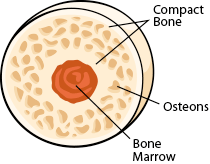
As the names suggest compact bone looks compact and the spongy bone looks like sponges.
This site is using cookies under cookie policy. Human tooth anatomy chalk painted. It suggests that the bone will have equal strength in all directions. The 2020 pandemic required that many of these activities be converted a format that students could complete online with a device, making labeling and coloring a little tricky. Sketch a typical long bone and label its epiphyses diaphysis. (a) anterior view with longitudinal endosteum yellow bone marrow compact bone periosteum perforating fibers nutrient arteries (c). Refer to as you study the following section. Anatomycorner is a branch of biologycorner.com focused on dissections and body systems. Two types of bone tissues in cross section of a long bone : As a nurse, you will need to know the basic about the human skeleton. Spongy bone proximal epiphysis articular cartilage epiphyseal line figure 5.2a the structure of a long bone (humerus). The compact bone is made up of osteon. The sloping roof of a building is a useful analogy to illustrate strike and dip.
The end of a growing tibia, cut lengthwise*. A = epiphysis b = diaphysis c = articular cartilage d = periosteum f = compact bone g = medullary cavity (yellow marrow) h = endosteum j = epiphyseal line (growth plate). Structure of long bone although there are many different types of bones in the skeleton, we will discuss the different parts of a specific type of bone epiphysis: The bottom sections of the spine are important when it comes to bearing weight and giving you a good center of gravity. It suggests that the bone will have equal strength in all directions.
6 3 Bone Structure Anatomy Physiology from open.oregonstate.education Two types of bone tissues in cross section of a long bone : Made up of small lumps of rocks with cracks and crevices. A vertical section through different layers of the soil is called the soil profile. Microscopic structure of a long bone. This quiz on human bones is designed to test your knowledge on the location of each individual bone. The end of a growing tibia, cut lengthwise*. As the names suggest compact bone looks compact and the spongy bone looks like sponges. The diaphysis of long bones mainly consists of compact bone, thus it offers bone strength.
A = epiphysis b = diaphysis c = articular cartilage d = periosteum f = compact bone g = medullary cavity (yellow marrow) h = endosteum j = epiphyseal line (growth plate).
Labeling portions of a long bone. The femur, the bone of the thigh, will be used as an example in considering the structure of a long bone. Observed 2.sketch and label the diaphysis of the beef bone: Geological cross sections are graphical representations of vertical slices through the earth used to clarify or interpret geological relationships with or without accompanying maps. The sloping roof of a building is a useful analogy to illustrate strike and dip. The structural unit of compact bone is osteon. rings of concentric lammelae (of canaliculi) are present around the central canal of each osteon. The end of a growing tibia, cut lengthwise*. Made up of small lumps of rocks with cracks and crevices. You can specify conditions of storing and accessing cookies in your browser. This site is using cookies under cookie policy. Refer to as you study the following section. The 2020 pandemic required that many of these activities be converted a format that students could complete online with a device, making labeling and coloring a little tricky. A = epiphysis b = diaphysis c = articular cartilage d = periosteum f = compact bone g = medullary cavity (yellow marrow) h = endosteum j = epiphyseal line (growth plate).